
How a Meniscus Tear Is Diagnosed
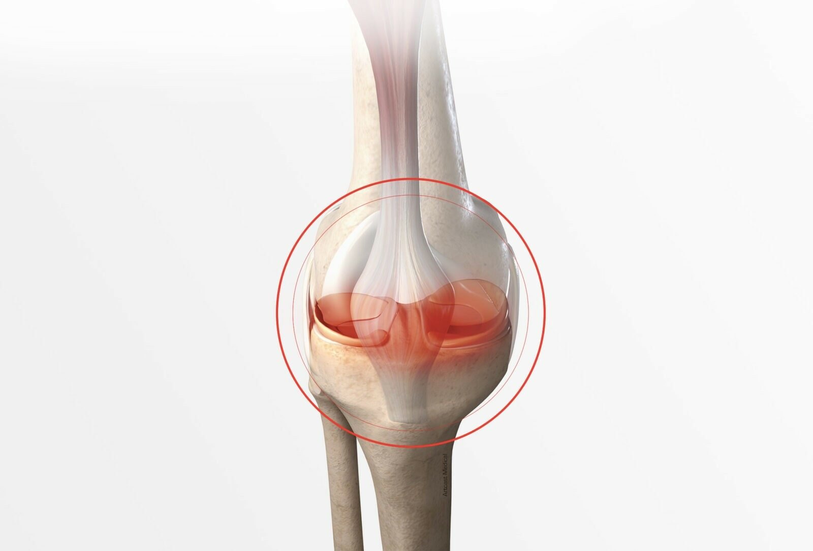
Being told you have a meniscus tear answers less than it appears to. Two knees with the same three words on the report can need completely different things: one needs stitches and four months, the other needs a rehabilitation programme and no surgery at all.
What separates them is not the diagnosis. It is where the tear sits, what shape it is, and how it got there. This guide covers how a meniscus tear is identified, what each test actually contributes, and which findings change the treatment.
What the meniscus does, and why losing it matters
Each knee has two menisci: C-shaped wedges of fibrocartilage between the femur and the tibia. They spread load across the joint, add stability, and protect the articular cartilage underneath.
That last function is the reason modern surgery tries to keep them. A knee that has lost meniscal tissue concentrates load on a smaller surface, and over years that accelerates arthritis. It is why our own approach is repair rather than removal wherever the tear allows it — the meniscus you keep is the protection your knee still has in twenty years.
How a meniscus tear usually presents
Two distinct stories, and they matter:
- The traumatic tear. A twist on a planted foot, often in sport. Immediate pain, swelling over the following day, and sometimes a sense that something moved inside the knee.
- The degenerative tear. No incident at all. A meniscus thinned by age splits under an ordinary movement — standing from a squat, getting out of a car. More common after forty-five, and frequently found alongside early arthritis.
Typical symptoms in both: pain along the joint line, swelling that comes and goes, clicking or catching, pain with twisting or deep bending, and difficulty fully straightening the knee.
Locking is the one to act on. A knee that physically will not straighten, or that jams intermittently, suggests a fragment displaced into the joint — a bucket-handle tear — and that is time-sensitive.
The examination comes first
The history does half the work
How it happened, how quickly it swelled, whether the knee locks or gives way, what movements reproduce the pain. A twisting injury in a twenty-five-year-old and a gradual ache in a fifty-five-year-old point to different tears before anyone has touched the knee.
Joint line tenderness
Pressing along the gap between femur and tibia, medially and laterally. It is the simplest meniscal sign and one of the more useful, though tenderness can also come from the ligament or the cartilage underneath.
The McMurray test
The knee is bent, then extended while the tibia is rotated, loading the meniscus against the joint surface. A palpable click with pain is a positive result.
The Thessaly test
You stand on the affected leg with the knee bent about twenty degrees and rotate your body over the fixed foot. It reproduces the loading pattern that provokes real-life symptoms, which is why many clinicians rate it highly.
What the examination cannot do
No single test is decisive. Each has meaningful false positives and false negatives, which is why they are used as a group and read alongside the history. A confident diagnosis from one positive click is a guess with a name attached.
Imaging: what each scan is actually for
X-ray
X-rays do not show the meniscus. They are taken to look at the bone and the joint space: fractures, loose bodies, and above all the degree of arthritis. Weight-bearing views matter, because a joint space that looks acceptable lying down can be bone on bone standing up. In a degenerative knee, the X-ray often changes the plan more than the MRI does.
MRI
The reference test for meniscal tissue: it shows the tear, its position, its pattern and the state of the surrounding cartilage and ligaments.
It also has a well-known trap. Meniscal tears appear on the MRIs of a large proportion of people over fifty who have no knee symptoms at all. Finding a tear on a scan does not prove it is the source of the pain — the clinical picture decides that, and an MRI ordered before an examination frequently sends treatment in the wrong direction.
Ultrasound
Occasionally used for the outer edge of the meniscus and for surrounding structures, but it cannot see inside the joint reliably. It is a supplement, not a substitute.
The part most guides skip: the type of tear decides the treatment
This is where a diagnosis becomes a plan.
Where the tear sits: the red zone and the white zone
The outer third of the meniscus has a blood supply — the “red zone”. The inner two-thirds has almost none — the “white zone”. A tear in the outer third can heal when it is stitched. The same tear near the centre usually cannot, because there is no blood supply to heal it.
That single anatomical fact is why one patient is offered a repair and another is not.
The patterns
- Longitudinal and bucket-handle. A tear along the length of the meniscus; when the fragment flips into the joint, the knee locks. Often in the vascular zone, and often repairable — these are the tears where prompt surgery genuinely matters.
- Radial. Cutting inwards from the free edge. It interrupts the circumferential fibres that let the meniscus spread load, so even a small radial tear can cost much of its function.
- Horizontal. Splitting the meniscus into upper and lower layers, typical of degenerative tears.
- Flap and complex. Mixed patterns, common in older knees, frequently alongside arthritis.
- Root tears. The meniscus detaches at its attachment to the bone. Biomechanically this behaves like losing the whole meniscus, and it is the pattern most often overlooked on a report read quickly. It is also the one where an early repair changes the long-term outlook most.
When surgery is not the answer
Plenty of meniscal tears are managed without an operation, and in some groups that is the better medicine:
- Degenerative tears with established arthritis. Here the tear is usually a symptom of the arthritic knee, not the cause of the pain. Trimming it rarely helps, and can accelerate wear.
- Small, stable tears that do not lock or catch, especially in the outer zone where healing is possible.
- Anyone who has not yet done a proper rehabilitation programme. Quadriceps and hip strengthening, load management and time resolve a significant proportion of meniscal symptoms.
The honest test is not the scan. It is whether the knee’s mechanical symptoms — locking, catching, giving way — persist after a well-run programme.
When it is, and which operation
Surgery earns its place when the knee locks, when a repairable tear is found in a young or active patient, when a root tear is identified, or when good rehabilitation has failed to settle a mechanically unstable knee.
Three approaches, all arthroscopic, all on our meniscus page:
- Repair with suture, US$ 11,300 — stitching the tear. The preferred option whenever the tear’s position and pattern allow it.
- Meniscal remodeling, US$ 11,300 — reshaping the torn portion when it cannot hold a repair, removing as little tissue as possible.
- Wrapping with a collagen matrix, US$ 15,200 — supporting healing in tears at the margin of what can be repaired.
Every figure is published on our price list, and the wider procedure is described on our knee arthroscopy page.
Recovery: a trim and a repair are not the same thing
This distinction is worth insisting on, because it is where expectations go wrong.
- After a partial removal or remodeling, most patients resume light activity within two to four weeks and full activity, sport included, within six to eight weeks with physical therapy.
- After a repair, the stitched tissue has to heal before it is loaded. That means protected weight-bearing and a restricted range at first, then a staged progression — typically several months before pivoting sport, not several weeks.
A repair asks more of you in the first year and gives more back over the next twenty. That trade is the whole argument for keeping the meniscus.
When to see a specialist promptly
- The knee locks or will not straighten fully
- It gives way under you
- Swelling that recurs after activity
- Pain that has not settled after four to six weeks of sensible self-management
- Any knee injury with immediate swelling within hours, which suggests bleeding in the joint
Frequently asked questions
Can a meniscus tear heal on its own?
A small tear in the outer, vascular zone can. Tears in the inner two-thirds generally cannot, because the blood supply that would heal them is not there. Symptoms settling is not the same as the tear healing.
Do I need an MRI to diagnose a meniscus tear?
Not always. History and examination often make the diagnosis, and in an older knee with clear arthritis the X-ray may matter more. An MRI is most useful when the result would change the plan — when a repair is being considered, or when the picture does not add up.
Is surgery always needed for a torn meniscus?
No, and for degenerative tears in arthritic knees it is often the wrong answer. Locking, a repairable tear in an active patient, and root tears are the situations where surgery is most clearly indicated.
What happens if I leave a meniscus tear untreated?
It depends on the type. A stable degenerative tear may cause no further harm. A bucket-handle fragment can damage the articular cartilage each time it displaces, and an untreated root tear behaves like a missing meniscus, which accelerates arthritis.
How long does the diagnosis itself take?
An examination and an X-ray can be done in one visit. An MRI adds days rather than weeks. The delay in most cases is not the testing; it is the months people spend deciding to be examined at all.
What to do next
If your knee locks, swells after activity, or has not settled in six weeks, the useful step is an examination and, if needed, imaging read by someone who will act on it. Send what you have — or tell us what the knee does — and a surgeon will tell you which tear pattern it looks like, whether it is repairable, and what that would cost, in writing, before you commit to anything.
You can also read how each knee procedure works on our knee surgery page.